
Dark Field Micros Pdf Microscopy Microscope Dark field microscopy is ideally used to illuminate unstained samples causing them to appear brightly lit against a dark background. this type of microscope contains a special condenser that scatters light and causes it to reflect off the specimen at an angle. This page titled 3.3a: dark field microscopy is shared under a cc by sa 4.0 license and was authored, remixed, and or curated by boundless via source content that was edited to the style and standards of the libretexts platform.
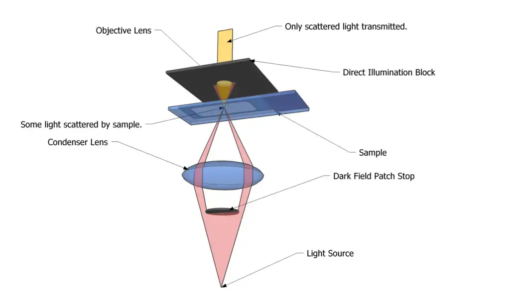
Dark Field Microscopy Principle Parts Procedure Uses Biology Two frequently used microscopy methods for seeing and evaluating materials are dark field microscopy and bright field microscopy. they differ in terms of lighting and picture generation, but they also have certain things in common. Dark field microscopy illuminates unstained samples against a dark background by using a hollow cone of light. the objective lens sits in the dark area and sees light refracted or scattered by the sample, appearing bright. it is used to view transparent or unstained specimens like microbes or cells. sample preparation is important to avoid background light scattering from dust or thickness. Chrome reader mode enter reader mode expand collapse global hierarchy home bookshelves microbiology microbiology (boundless) 3: microscopy. 3.1: looking at microbes 3.1a: microbe size 3.1b: units of measurement for microbes 3.1c: refraction and magnification 3.1d: magnification and resolution 3.2: other types of microscopy 3.2a: microscopy 3.2b: general staining methods 3.3: other types of microscopy 3.3a: dark field microscopy.
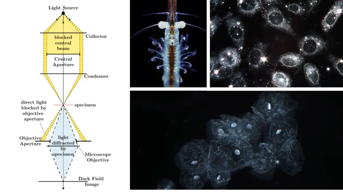
Dark Field Microscopy Principle Parts Procedure Uses Biology Chrome reader mode enter reader mode expand collapse global hierarchy home bookshelves microbiology microbiology (boundless) 3: microscopy. 3.1: looking at microbes 3.1a: microbe size 3.1b: units of measurement for microbes 3.1c: refraction and magnification 3.1d: magnification and resolution 3.2: other types of microscopy 3.2a: microscopy 3.2b: general staining methods 3.3: other types of microscopy 3.3a: dark field microscopy. Dark field microscopy worked by: dea tikvina afeza krakuli course: microbiology study program: biotechnology i fdark field microscopy • dark field microscopy is like peering into a hidden world illuminated by scattered light. • it's a technique where the specimen appears bright against a dark background, revealing intricate details that might otherwise remain unseen. • it's a window into. The 20th century saw the development of microscopes that leveraged nonvisible light, such as fluorescence microscopy, which uses an ultraviolet light source, and electron microscopy, which uses short wavelength electron beams. these advances led to major improvements in magnification, resolution, and contrast.
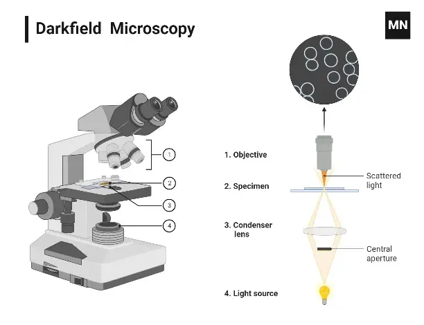
Dark Field Microscopy Principle Parts Procedure Uses Biology Dark field microscopy worked by: dea tikvina afeza krakuli course: microbiology study program: biotechnology i fdark field microscopy • dark field microscopy is like peering into a hidden world illuminated by scattered light. • it's a technique where the specimen appears bright against a dark background, revealing intricate details that might otherwise remain unseen. • it's a window into. The 20th century saw the development of microscopes that leveraged nonvisible light, such as fluorescence microscopy, which uses an ultraviolet light source, and electron microscopy, which uses short wavelength electron beams. these advances led to major improvements in magnification, resolution, and contrast.
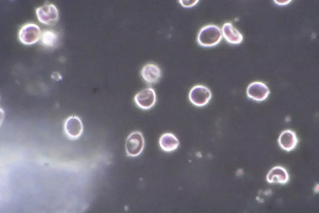
Dark Field Microscopy Principle Parts Procedure Uses Biology
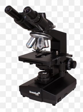
Dark Field Microscopy Images Dark Field Microscopy Transparent Png