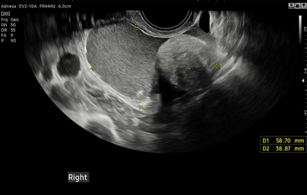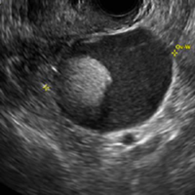
Dermoid Ovarian Cyst Ultrasound An unusual sonographic appearance of a dermoid cyst in a young patient a dermoid cyst is a rare benign cystic teratoma. on ultrasound, it has variable sonographic features since it is usually filled with an admixture of ectoderm, mesoderm, and endoderm origin contents. because of the lumen of the dermoid cyst is usually filled with an admixture of ectoderm, mesoderm and endoderm origin. Abstract ovarian dermoid cysts are made up of solid, cystic and fat tissue. these components give rise to characteristic sonographic features such as a fat‐fluid level, dermoid mesh and tip of the iceberg sign. the presence of two or more of these typical features can be used to confidently diagnose a dermoid cyst on ultrasound.

Dermoid Ovarian Cyst Ultrasound Ultrasound ultrasound is the preferred imaging modality. typically an ovarian dermoid is seen as a cystic adnexal mass with some mural components. most lesions are unilocular. the spectrum of sonographic features includes: diffusely or partially echogenic mass with posterior sound attenuation owing to sebaceous material and hair within the cyst. A dermoid cyst is a rare benign cystic teratoma. on ultrasound, it has variable sonographic features since it is usually filled with an admixture of ectoderm, mesoderm, and endoderm origin contents. Yet, a thorough analysis of all ultrasound features that characterize dermoid cysts can lead in the vast majority of the cases to an exact diagnosis. the purpose of this paper is to present the ultra sonographic fi ndings of the dermoid cysts. key words: ovarian dermoid cyst, mature cystic teratoma, ultrasonography. On ultrasound, dermoid cysts typically present as well defined, round or oval masses within the ovary. one of their hallmark features is the presence of a mixture of different tissue types, which can create a distinctive appearance.

Dermoid Ovarian Cyst Ultrasound Yet, a thorough analysis of all ultrasound features that characterize dermoid cysts can lead in the vast majority of the cases to an exact diagnosis. the purpose of this paper is to present the ultra sonographic fi ndings of the dermoid cysts. key words: ovarian dermoid cyst, mature cystic teratoma, ultrasonography. On ultrasound, dermoid cysts typically present as well defined, round or oval masses within the ovary. one of their hallmark features is the presence of a mixture of different tissue types, which can create a distinctive appearance. Ultrasound of ovarian dermoids – sonographic findings of a dermoid cyst in a 41 year old woman with an elevated serum hcg. australasian journal of ultrasound in medicine. Dermoid cyst is the most common serm cell tumor of ovary, comprise approximately 20% of all ovarian tumors. however on gross appearance cyst containing multiple fat balls of varying size is very rare.

Dermoid Ovarian Cyst Ultrasound Ultrasound of ovarian dermoids – sonographic findings of a dermoid cyst in a 41 year old woman with an elevated serum hcg. australasian journal of ultrasound in medicine. Dermoid cyst is the most common serm cell tumor of ovary, comprise approximately 20% of all ovarian tumors. however on gross appearance cyst containing multiple fat balls of varying size is very rare.

Dermoid Cyst Ultrasound Case 164 57 Off