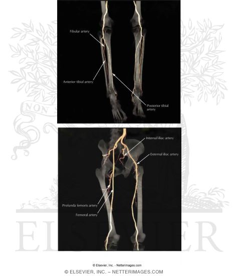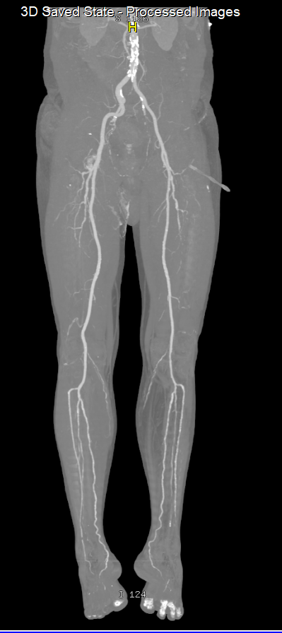
Lower Limb Arteries Lower Limb Ct Angiography Anatomy Radiology Hot An example of normal arterial vascular anatomy (with some areas of intimal disease) of the lower limb. This angiographic atlas of arteries of the lower limb has been designed to help radiologists and vascular surgeons in their daily practice. lower limb computed tomography angiography (cta) is a valuable noninvasive imaging modality used primarily for the diagnosis and management of peripheral arterial disease (pad) and acute limb ischemia (ali).

Arteries Of Lower Limb Pdf Human Leg Lower Limb Anatomy This video explains the lower limb #arterial #anatomy as seen in #ct scan. The arterial supply to the lower limb is chiefly supplied by the femoral artery and its branches. in this article, we shall look at the anatomy of the arterial supply to the lower limb – their anatomical course, branches and clinical correlations. This document provides information on performing and interpreting ct angiography of the lower limbs. it discusses scanning techniques, protocols, contrast injection, and principles of timing acquisitions. image post processing includes mip, vr, and mpr. interpretation requires scrutinizing calcifications and stents to avoid overestimating stenosis. peripheral cta is useful for evaluating. Optimized visualization of peripheral ct angiography datasets using bone removal. (a) coronal volume rendered images from a ct angiogram of the lower extremities obtained in a 63 year old male with loeys dietz syndrome demonstrate ectatic and tortuous superficial femoral arteries, however, the popliteal arteries and calf vessels are not visualized due to overlying bone. bone subtracted coronal.

Ct Angiograms Of Arteries Of The Lower Limb This document provides information on performing and interpreting ct angiography of the lower limbs. it discusses scanning techniques, protocols, contrast injection, and principles of timing acquisitions. image post processing includes mip, vr, and mpr. interpretation requires scrutinizing calcifications and stents to avoid overestimating stenosis. peripheral cta is useful for evaluating. Optimized visualization of peripheral ct angiography datasets using bone removal. (a) coronal volume rendered images from a ct angiogram of the lower extremities obtained in a 63 year old male with loeys dietz syndrome demonstrate ectatic and tortuous superficial femoral arteries, however, the popliteal arteries and calf vessels are not visualized due to overlying bone. bone subtracted coronal. Anatomy included indications (1,2) lower extremity trauma high risk injuries include – distal segmental femoral fractures, high energy tibial plateau fractures, displaced distal femoral fractures at the adductor hiatus, open femoral or tibial fractures, knee dislocation. arterial laceration, occlusion, dissection or av fistula aneurysm. Computed tomographic angiography (cta) can provide more detailed overview of the lower limb vasculature, rule out or confirm an uncertain pad diagnosis or verify the degree and an exact location of the stenosis prior to the revascularization [3 – 5].

Computed Tomography Ct Angiography Of Bi Lateral Lower Limb Anatomy included indications (1,2) lower extremity trauma high risk injuries include – distal segmental femoral fractures, high energy tibial plateau fractures, displaced distal femoral fractures at the adductor hiatus, open femoral or tibial fractures, knee dislocation. arterial laceration, occlusion, dissection or av fistula aneurysm. Computed tomographic angiography (cta) can provide more detailed overview of the lower limb vasculature, rule out or confirm an uncertain pad diagnosis or verify the degree and an exact location of the stenosis prior to the revascularization [3 – 5].

Computed Tomography Ct Angiography Of Bi Lateral Lower Limb

Best Ct Scan Lower Limb Angiography In Pune Nanded City