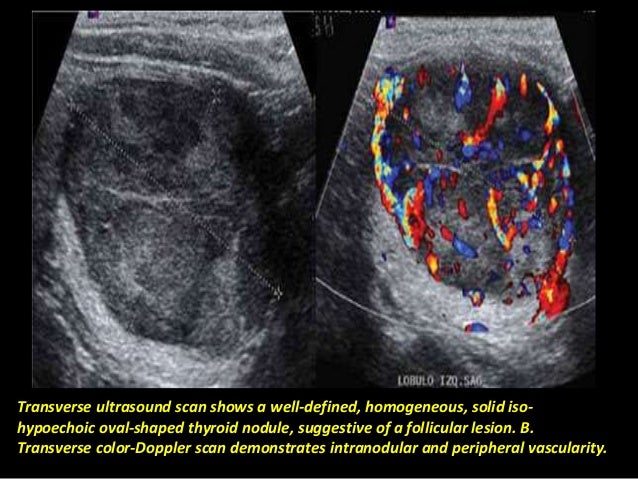
Papillary Thyroid Carcinoma Pdf Hyperthyroidism Glands Color doppler ultrasound: provides insights into blood flow, where patterns like “spoke wheel” could signify papillary thyroid carcinoma. by carefully analyzing these ultrasound images, doctors can differentiate between benign and potentially malignant nodules, ultimately improving diagnosis and treatment plans. Papillary thyroid carcinoma is a form of cancer that occurs due to abnormal and uncontrolled cell growth of certain cells follicular cells of the thyroid. papillary thyroid carcinoma ptc arising within a follicular adenoma is an exceptionally rare histopathological subtype that shows the nuclear features of ptc within a benign appearing.

Papillary Thyroid Cancer Ultrasound Colors Warehouse Of Ideas Papillary thyroid cancer (as is the case with follicular thyroid cancer) typically occurs in the middle aged, with a peak incidence in the 3 rd and 4 th decades. Use of ultrasound in thyroid cancer imaging equipment and technique an advanced ultrasound machine with a high frequency transducer (7.5–12 mhz) is the basic equipment required. high frequency transducers allow superior near field resolution and form the basis of characterization of benign and malignant thyroid nodules. After surgical removal of the thyroid gland or part of it due to papillary thyroid cancer, ultrasound is routinely employed to assess the surgical bed and surrounding lymph nodes in the neck. this ongoing monitoring helps detect any potential recurrence of the cancer or the development of new suspicious nodules. Longitudinal color doppler ultrasound image shows increased central and peripheral vascularity in a follicular variant of papillary carcinoma thyroid nodule in a 14 year old male. rsna photo incidental contrast enhanced ct image of thyroid nodule in a 62 year old man with history of lymphoma involving the parotid gland.

Papillary Thyroid Cancer Ultrasound Colors Warehouse Of Ideas After surgical removal of the thyroid gland or part of it due to papillary thyroid cancer, ultrasound is routinely employed to assess the surgical bed and surrounding lymph nodes in the neck. this ongoing monitoring helps detect any potential recurrence of the cancer or the development of new suspicious nodules. Longitudinal color doppler ultrasound image shows increased central and peripheral vascularity in a follicular variant of papillary carcinoma thyroid nodule in a 14 year old male. rsna photo incidental contrast enhanced ct image of thyroid nodule in a 62 year old man with history of lymphoma involving the parotid gland. Papillary carcinoma of the thyroid typically appears as a solid, hypoechoic nodule with irregular or microlobulated margins on ultrasound. it often shows a t. Thyroid ultrasound is the best imaging test to diagnose and evaluate papillary thyroid cancer. when there is concern or suspicion for papillary thyroid cancer, a skilled, high resolution ultrasound of the thyroid, surrounding structures, and lymph nodes in the neck should be done first (not a ct scan or mri).

Papillary Thyroid Cancer Ultrasound Colors Warehouse Of Ideas Papillary carcinoma of the thyroid typically appears as a solid, hypoechoic nodule with irregular or microlobulated margins on ultrasound. it often shows a t. Thyroid ultrasound is the best imaging test to diagnose and evaluate papillary thyroid cancer. when there is concern or suspicion for papillary thyroid cancer, a skilled, high resolution ultrasound of the thyroid, surrounding structures, and lymph nodes in the neck should be done first (not a ct scan or mri).

Papillary Thyroid Cancer Ultrasound Colors Warehouse Of Ideas