Pelvis Xray Diagram Quizlet Being systematical is crucial to make possible for the non specialist to interpret pelvic radiographs accurately. table 1 shows the summary of how to read a pelvic x ray. references and further reading lee c, porter k. the prehospital management of pelvic fractures. emergency medicine journal: emj. 2007 feb;24 (2):130 3. pubmed pmid: 17251627. Pelvis xray important lines on a pelvis xray and how to read them navendu goyal 2.21k subscribers subscribed.

X Ray Pelvis Pdf The pelvis has complex anatomic structures that form the basis of the lines, arcs and stripes concept in the assessment of pelvic radiographs. having good knowledge of the pelvic anatomy and the spectrum of diseases is an important prerequisite for making an accurate diagnosis. The pelvis radiograph is comprised of the innominate hip bones or os coxae (ischium, pubis and ilium), the sacrum and the proximal femur. much of the interpretation is down to regions, rings and lines alongside an understanding of traumatic fracture patterns of the pelvic ring. Introduction interpreting pelvic x rays is a crucial skill in many medical and healthcare fields as it provides vital information for diagnosing and treating patients. this guide aims to introduce readers to pelvic x rays and provide three essential methods to interpret them. understanding pelvic x rays (pxrs) a pelvic x ray is a common imaging test that captures images of the pelvic bones. No pelvic tilt coccyx located 2cm above pubic symphysis no rotation of pelvis sacrum in midline symmetrical greater trochanters obturator foramen no visualization of lesser trochanters too much external rotation of leg leads to increased visualization of lesser trochanter if lesser trochanters are visible, they should be of symmetrical size.

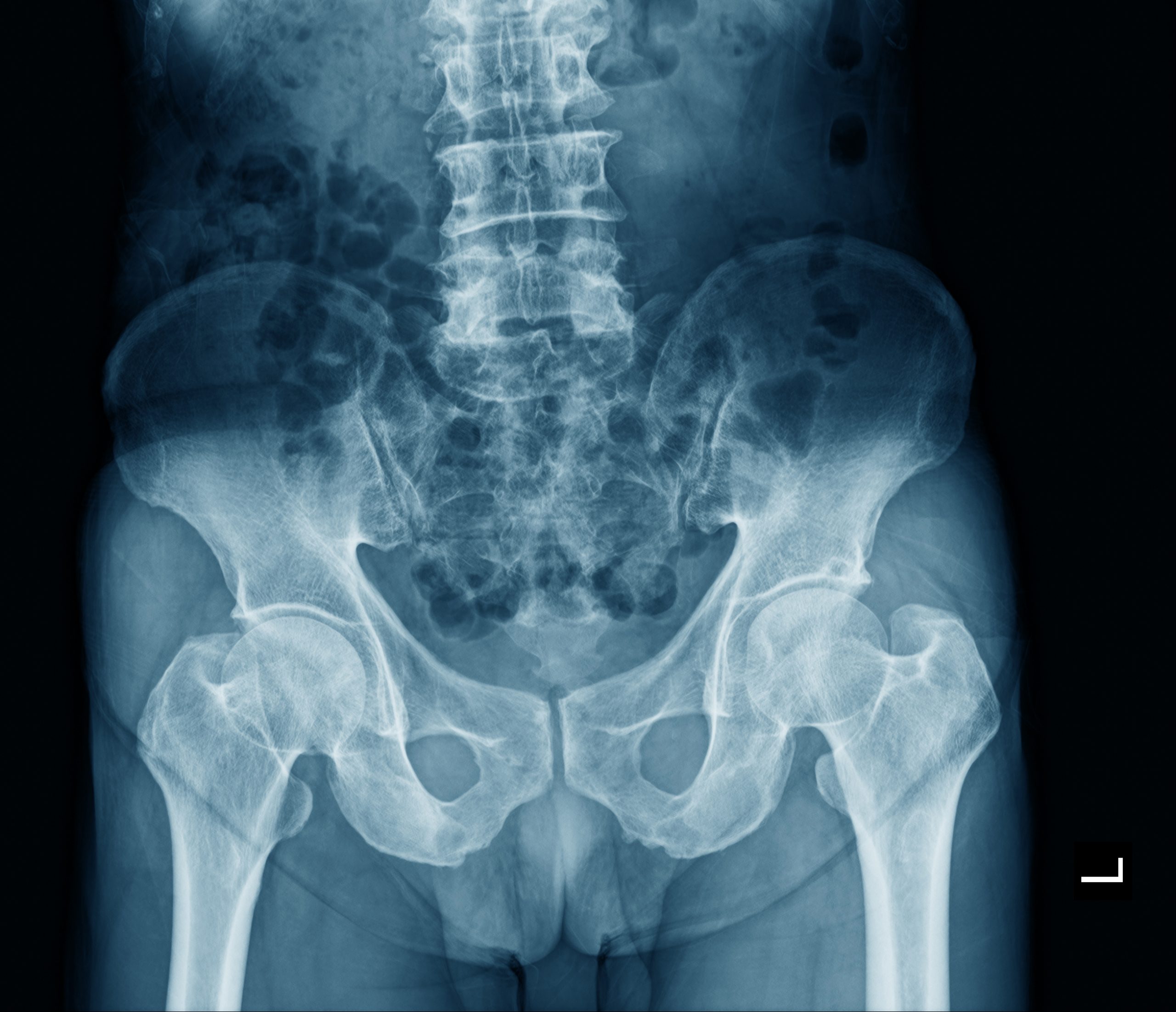
Xray Of Pelvis Diagram Quizlet Introduction interpreting pelvic x rays is a crucial skill in many medical and healthcare fields as it provides vital information for diagnosing and treating patients. this guide aims to introduce readers to pelvic x rays and provide three essential methods to interpret them. understanding pelvic x rays (pxrs) a pelvic x ray is a common imaging test that captures images of the pelvic bones. No pelvic tilt coccyx located 2cm above pubic symphysis no rotation of pelvis sacrum in midline symmetrical greater trochanters obturator foramen no visualization of lesser trochanters too much external rotation of leg leads to increased visualization of lesser trochanter if lesser trochanters are visible, they should be of symmetrical size. Views ap pelvis: patient supine and the x ray beam oriented 90 degrees to the patient’s long axis, passing through the patient from anterior to posteriorpubic symphysis and coccyx in straight line in middle of screen. Example: this is an ap pelvis in a skeletally mature individual, and there are no apparent fractures, some mild osteoarthritis in hip joints present. next, after you have stated what you can from quick glance it is a good practice to go through the whole image systematically. system for reading pelvis xrays check for correct patient and image date check quality of image spinous processes.

Xray Of Pelvis Diagram Quizlet Views ap pelvis: patient supine and the x ray beam oriented 90 degrees to the patient’s long axis, passing through the patient from anterior to posteriorpubic symphysis and coccyx in straight line in middle of screen. Example: this is an ap pelvis in a skeletally mature individual, and there are no apparent fractures, some mild osteoarthritis in hip joints present. next, after you have stated what you can from quick glance it is a good practice to go through the whole image systematically. system for reading pelvis xrays check for correct patient and image date check quality of image spinous processes.

Pelvis X Ray Anatomical Evaluation Imaging Stock Photo 2502786917

Radiographic Assessment Pelvis X Ray Analysis Stock Photo 2502786911

Pelvis X Ray Aarthi Scans And Labs