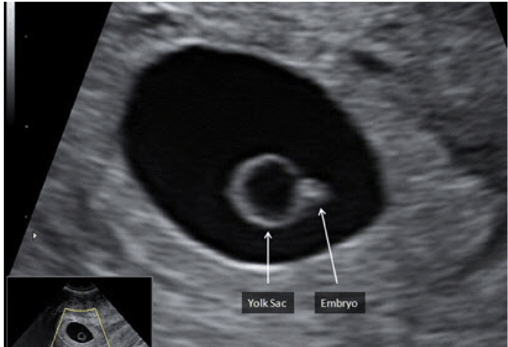
5 Weeks Gestational Sac And Yolk Sac But No Fetal Pole Yet Is That By five weeks gestation, we are likely to see at least a gestational sac. with transvaginal ultrasound, an intrauterine pregnancy can usually be seen with a beta hcg of 1,000 1,500 iu l. sometimes we can also see a yolk sac and by about 5w5d, we may even see a fetal pole with cardiac motion. Yes this happened to me exactly on 5 6, right down to the very faint possible but not definite yolk sac. when i went back at 6 3 there was a fetal pole with heartbeat. baby was born recently.

6 Weeks Ultrasound No Yolk Sac Or Fetal Pole Babycenter At 5 weeks, your baby is only about the size of a peppercorn. the only things you’re likely to see on an ultrasound are the yolk sac and the gestational sac. and even those may not be visible. At this point, an early heartbeat may be detected using high resolution transvaginal ultrasound, although it’s still possible for it to be too early depending on implantation timing and equipment quality. not seeing the fetal pole or heartbeat at exactly six weeks doesn’t necessarily mean there’s a problem. Hey all, ivf pregnancy here. went in for my first transvaginal ultrasound last week and they found the gestational and yolk sacs measuring 6 weeks but no fetal pole. The timing of events in early pregnancy — gestational sac at 5 weeks, yolk sac at 5 ½ weeks, and embryo with heartbeat at 6 weeks — is accurate and reproducible, with a variation of about ± ½ week; this consistency explains the time related criteria for pregnancy failure.

Yolk Sac Fetal Pole Hey all, ivf pregnancy here. went in for my first transvaginal ultrasound last week and they found the gestational and yolk sacs measuring 6 weeks but no fetal pole. The timing of events in early pregnancy — gestational sac at 5 weeks, yolk sac at 5 ½ weeks, and embryo with heartbeat at 6 weeks — is accurate and reproducible, with a variation of about ± ½ week; this consistency explains the time related criteria for pregnancy failure. A mass of fetal cells, separate from the yolk sac, first becomes apparent on transvaginal ultrasound just after the 6th week of gestation. this mass of cells is known as the fetal pole. The embryo is initially visualized as an echogenic area of thickening on the yolk sac. straight until it reaches 3 4mm in length (on ultrasound the embryo is a straight echogenic line adjacent to the yolk sac and close to the vitelline duct).

Yolk Sac Fetal Pole Ob Images A mass of fetal cells, separate from the yolk sac, first becomes apparent on transvaginal ultrasound just after the 6th week of gestation. this mass of cells is known as the fetal pole. The embryo is initially visualized as an echogenic area of thickening on the yolk sac. straight until it reaches 3 4mm in length (on ultrasound the embryo is a straight echogenic line adjacent to the yolk sac and close to the vitelline duct).