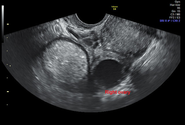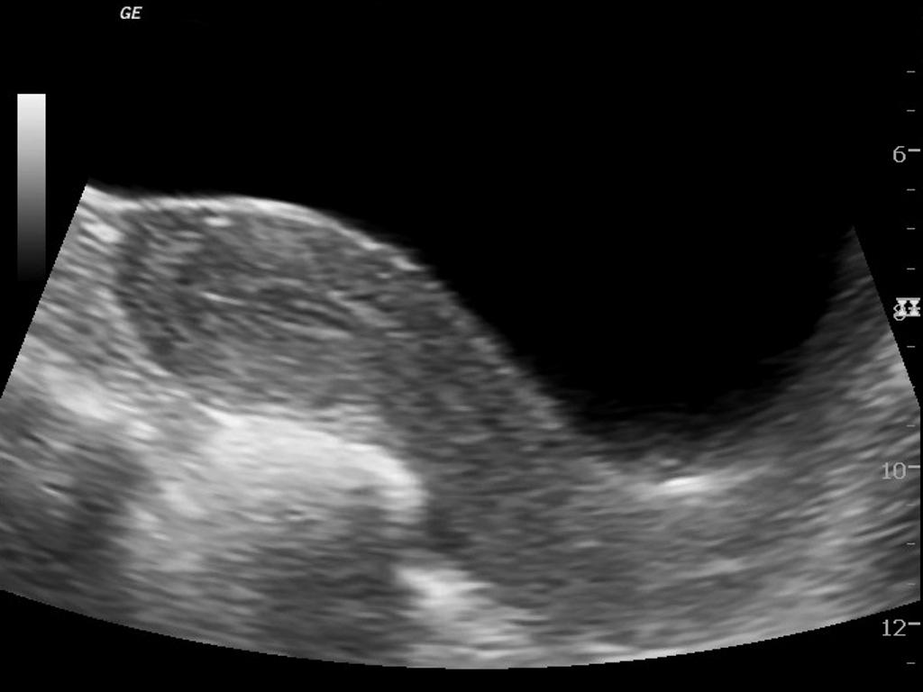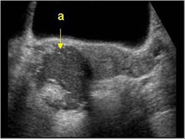
Dermoid Cyst Ultrasound Case 164 57 Off In this radiology lecture, we discuss the ultrasound appearance of ovarian dermoid cyst, including the rarely seen but highly specific “floating sphere” sign!. Dermoid cyst || ultrasound || case 164 clinical features: a 19 years old female patient came with lower abdominal pain. ultrasound features: a well defined, round to oval, sound attenuating.

Dermoid Cyst Ultrasound Wikidoc Dr tahir a siddiqui ( consultant sonologist )ultrasound learning video seriesgujranwala. pakistan0553257350. Bilateral ovarian dermoid cyst | treatment | complications | ultrasound kashif ultrasound made easy cases 8.92k subscribers subscribed. @uniqueradiologist99 right ovarian dermoid cyst #ultrasound #doctor 柔和之光 · 萧萧. Dermoid cysts contain skin elements, including squamous epithelium and dermal appendages (adnexa), such as sebaceous and sweat glands, and hair. they are discussed separately according to anatomic location: intracranial dermoid cyst orbital de.

Dermoid Ovarian Cyst Ultrasound @uniqueradiologist99 right ovarian dermoid cyst #ultrasound #doctor 柔和之光 · 萧萧. Dermoid cysts contain skin elements, including squamous epithelium and dermal appendages (adnexa), such as sebaceous and sweat glands, and hair. they are discussed separately according to anatomic location: intracranial dermoid cyst orbital de. Abstract ovarian dermoid cysts are made up of solid, cystic and fat tissue. these components give rise to characteristic sonographic features such as a fat‐fluid level, dermoid mesh and tip of the iceberg sign. the presence of two or more of these typical features can be used to confidently diagnose a dermoid cyst on ultrasound. The initial ultrasound scan showed the characteristic sonographic features of an ovarian dermoid cyst. ct abdomen was done later which confirmed the findings. this case highlights some of the classic features of an ovarian dermoid cyst viz. rokitansky nodule, tip of iceberg sign, linear mesh of hair, fat fluid levels and tooth ossification within.

Dermoid Ovarian Cyst Ultrasound Abstract ovarian dermoid cysts are made up of solid, cystic and fat tissue. these components give rise to characteristic sonographic features such as a fat‐fluid level, dermoid mesh and tip of the iceberg sign. the presence of two or more of these typical features can be used to confidently diagnose a dermoid cyst on ultrasound. The initial ultrasound scan showed the characteristic sonographic features of an ovarian dermoid cyst. ct abdomen was done later which confirmed the findings. this case highlights some of the classic features of an ovarian dermoid cyst viz. rokitansky nodule, tip of iceberg sign, linear mesh of hair, fat fluid levels and tooth ossification within.