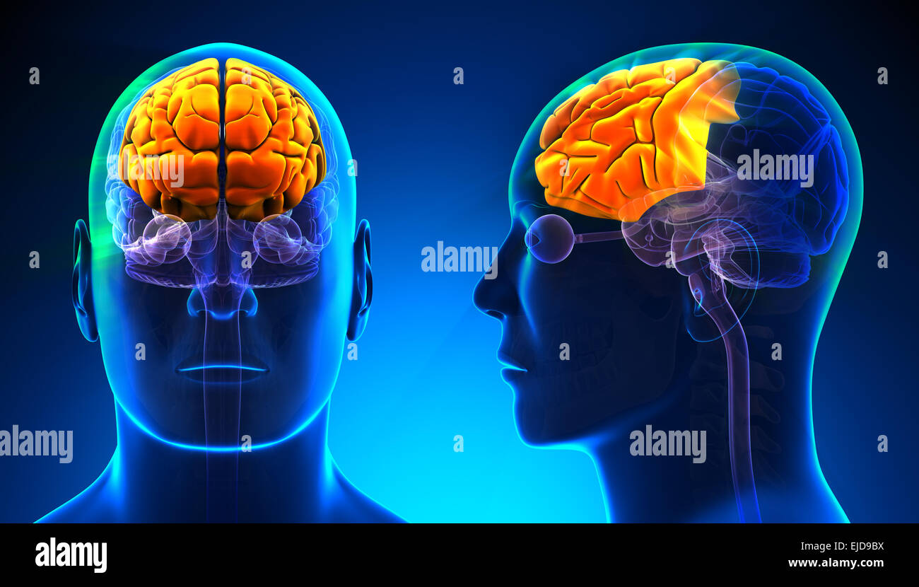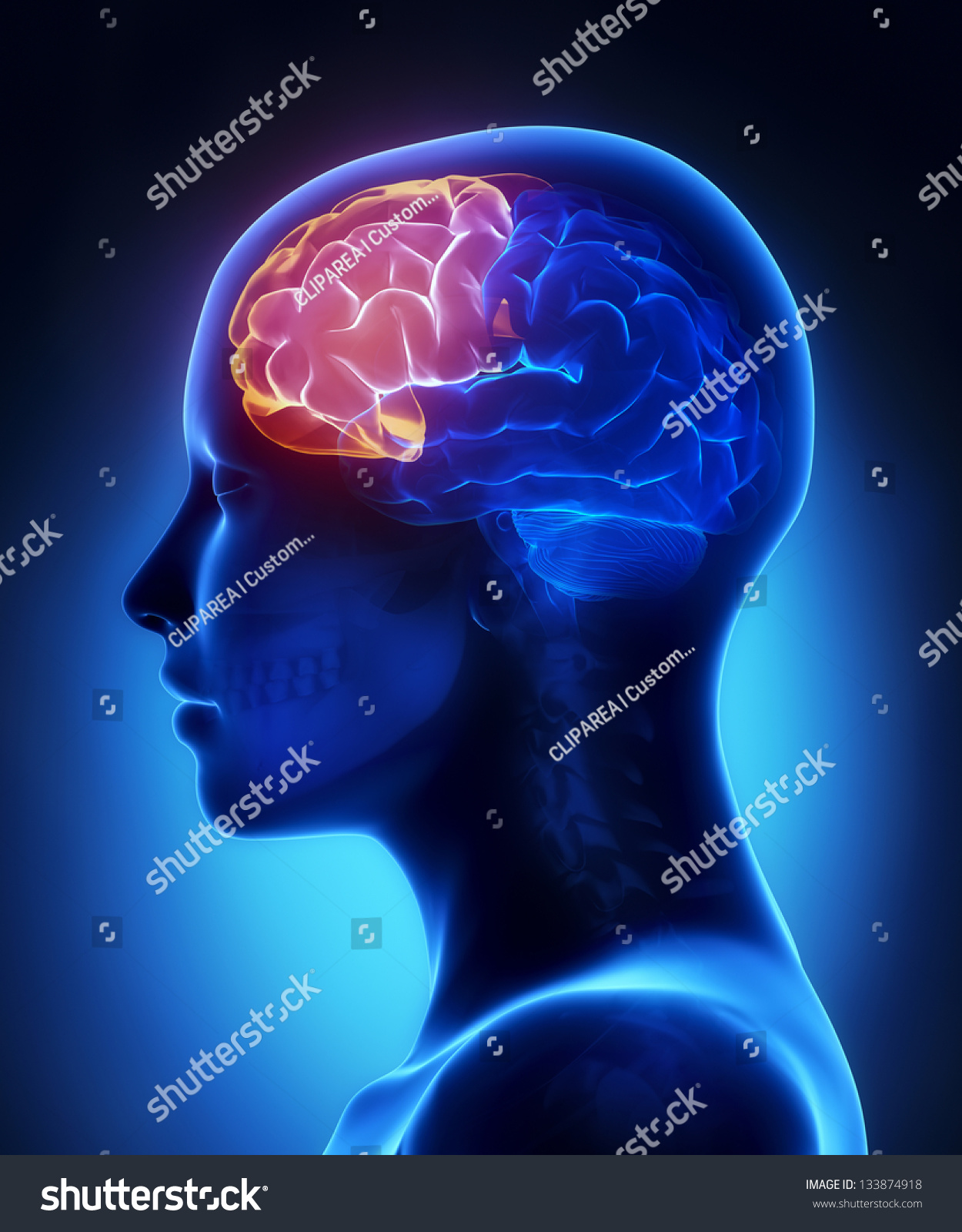
Human Brain Frontal Lobe Anatomy Human Brain Anatomy 3d 55 Off Your brain’s frontal lobe is home to areas that manage thinking, emotions, personality, judgment, self control, muscle control and movements, memory storage and more. just as its name indicates, it’s the forward most area of your brain. your frontal lobe is a key area of study for both brain related and mental health related fields of medicine. Parietal lobe function and left frontal lobe function the parietal lobe is located at the top and back of the brain. it plays a critical role in processing sensory information and spatial awareness. the left frontal lobe is responsible for language production, logical thinking, and problem solving.

Frontal Lobe Brain Anatomy The four lobes in your brain are called the frontal, parietal, temporal, and occipital lobes. each of these lobes controls specific functions of your body. The frontal lobe damage can cause a range of symptoms related to decision making, physical movements, and self control. frontal lobe damage impairs quality of life. The frontal lobe is the largest of the four major lobes of the brain in mammals, and is located at the front of each cerebral hemisphere (in front of the parietal lobe and the temporal lobe). it is parted from the parietal lobe by a groove between tissues called the central sulcus and from the temporal lobe by a deeper groove called the lateral sulcus (sylvian fissure). the most anterior. The cerebral cortex is the outermost portion of the brain, containing billions of tightly packed neuronal cell bodies that form what is known as the gray matter of the brain. the white matter of the brain and spinal cord forms as heavily myelinated axonal projections of these neuronal cell bodies. the cerebral cortex has four major divisions known as lobes: the frontal, temporal, parietal, and.

Frontal Lobe Brain Anatomy The frontal lobe is the largest of the four major lobes of the brain in mammals, and is located at the front of each cerebral hemisphere (in front of the parietal lobe and the temporal lobe). it is parted from the parietal lobe by a groove between tissues called the central sulcus and from the temporal lobe by a deeper groove called the lateral sulcus (sylvian fissure). the most anterior. The cerebral cortex is the outermost portion of the brain, containing billions of tightly packed neuronal cell bodies that form what is known as the gray matter of the brain. the white matter of the brain and spinal cord forms as heavily myelinated axonal projections of these neuronal cell bodies. the cerebral cortex has four major divisions known as lobes: the frontal, temporal, parietal, and. The frontal lobe is the largest lobe of the brain, occupying about one third of the cerebral hemisphere. as the name implies, the frontal lobe is located in the anterior aspect of the cranial cavity, conforming to the inner surface of the frontal bone. the frontal lobe is separated from the parietal lobe posteriorly by a groove called the central sulcus, and from the temporal lobe. Clinically, frontal lobe syndromes, frontal network syndromes, frontal systems syndromes, executive dysfunction, and metacognition have all been used to describe disorders of frontal lobes and their extended networks although they are not all synonymous. anatomically they refer to those parts of the brain rostral to the central sulcus.

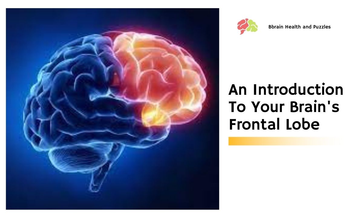
An Introduction To Your Brain S Frontal Lobe Brain Health And Puzzles The frontal lobe is the largest lobe of the brain, occupying about one third of the cerebral hemisphere. as the name implies, the frontal lobe is located in the anterior aspect of the cranial cavity, conforming to the inner surface of the frontal bone. the frontal lobe is separated from the parietal lobe posteriorly by a groove called the central sulcus, and from the temporal lobe. Clinically, frontal lobe syndromes, frontal network syndromes, frontal systems syndromes, executive dysfunction, and metacognition have all been used to describe disorders of frontal lobes and their extended networks although they are not all synonymous. anatomically they refer to those parts of the brain rostral to the central sulcus.

668 Frontal Lobe Brain Stock Photos High Res Pictures And Images