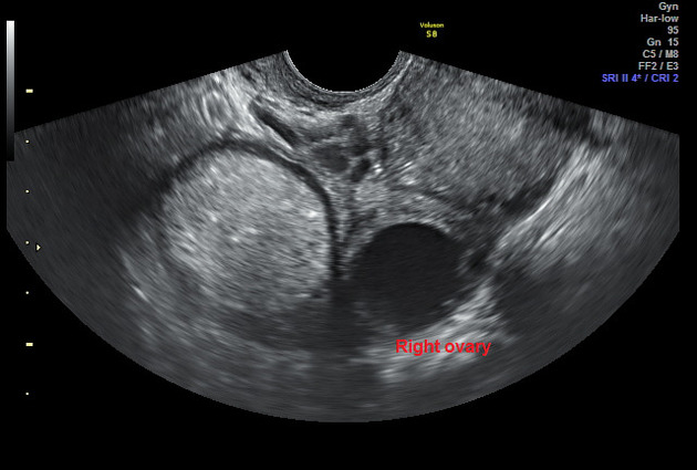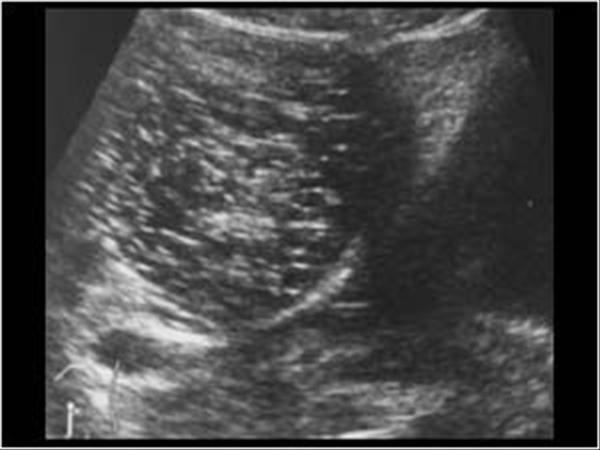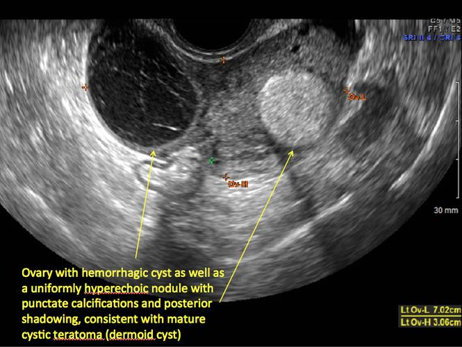
Bilateral Simple Cysts Ovaries By Ultrasound Stock Photo 1757857103 Both ovaries show small dermoid cysts. the dermoid cyst is seen as a small hyperechoic lesion in the ovary. see: ultrasound images ovaries.htm#. Both ovaries reveal complex multilocular cysts within. these locules reveal fat content (hyperintense on t1w and signal loss on fat suppressed sequences) suggesting dermoid cysts. the right ovarian dermoid cyst measures about 4.5 x 4 x 3.8 cm in its maximum dimensions.

Dermoid Cyst Ultrasound Case 164 57 Off In summary, bilateral ovarian dermoid cysts are generally benign but may require medical or surgical intervention depending on size, symptoms, and potential complications. regular gynecological checkups and imaging play a key role in early detection and effective management. A non septated peritoneal pseudocyst with the ovary seen separately containing an endometrioma and follicles in the cortex. the patient has a clinical history of multiple surgical procedures for endometriosis. paraovarian cysts paraovarian cysts arise in the broad ligament between the ovary and the fallopian tube. they account for 5–20% of adnexal masses (19, 20). the incidence of borderline. Shown are transvaginal ultrasound images of two patients that demonstrate the 'tip of the iceberg' sign: acoustic shadowing from the hyperechoic part of the dermoid cyst (arrow). Cumulus oophorus functional cysts of the ovary a) follicular cysts this young female patient underwent sonography for non specific pain in the lower abdomen. ultrasound images of the pelvis show bilateral ovarian cysts which show absence of internal nodules, septae or debris. these findings are typical of follicular cysts of the ovaries.

Ultrasound Imaging Of Ovarian Dermoid Cysts Shown are transvaginal ultrasound images of two patients that demonstrate the 'tip of the iceberg' sign: acoustic shadowing from the hyperechoic part of the dermoid cyst (arrow). Cumulus oophorus functional cysts of the ovary a) follicular cysts this young female patient underwent sonography for non specific pain in the lower abdomen. ultrasound images of the pelvis show bilateral ovarian cysts which show absence of internal nodules, septae or debris. these findings are typical of follicular cysts of the ovaries. Ultrasound is an essential tool in the diagnosis and management of ovarian cysts, providing detailed images that help determine the type and size of the cysts. with its ability to distinguish between simple, complex, and potentially malignant cysts, ultrasound ensures timely and accurate diagnosis, allowing for appropriate treatment decisions. Ultrasound of ovarian dermoid cyst in this radiology lecture, we discuss the ultrasound appearance of ovarian dermoid cyst, including the rarely seen but highly specific “floating sphere” sign! key points include aka mature cystic teratoma. most common ovarian neoplasm. benign, mean age 30. 10% bilateral.

Ultrasound Image Of Bilateral Ovarian Dermoid Cyst Download Ultrasound is an essential tool in the diagnosis and management of ovarian cysts, providing detailed images that help determine the type and size of the cysts. with its ability to distinguish between simple, complex, and potentially malignant cysts, ultrasound ensures timely and accurate diagnosis, allowing for appropriate treatment decisions. Ultrasound of ovarian dermoid cyst in this radiology lecture, we discuss the ultrasound appearance of ovarian dermoid cyst, including the rarely seen but highly specific “floating sphere” sign! key points include aka mature cystic teratoma. most common ovarian neoplasm. benign, mean age 30. 10% bilateral.

Ultrasound Image Of Bilateral Ovarian Dermoid Cyst Download

Dermoid Ovary Ultrasound

Dermoid Cysts On Tumblr